Helminthiasis in pets is a completely common thing; all more or less experienced breeders and pet lovers have encountered them. But what association do you have from the word “worms”? You probably immediately remember the worms that live in the digestive tract of animals. Of course, this is not without reason: this is where the vast majority of parasites are localized. But don’t think that all worms are so “banal.” There are, for example, even subcutaneous worms in dogs!
Basic information
It is interesting that the term “subcutaneous” can mean several types of parasites , but we will start with the most unusual and severe option. There are helminths from the genus Dirofilaria that cause a disease called dirofilariasis. It is also called “heartworm”. Yes, under normal conditions these worms parasitize in the lumen of the pulmonary artery, aorta, or directly in the cavity of the ventricles/atria. The disease in dogs is severe, and deaths are common. The pathology is especially widespread in the United States and Canada, and the Great Lakes region was notorious among the Indians: worms can also parasitize the human body.
A small digression from the topic: initially, people did not suffer from dirofilariasis. More precisely, infection could have occurred, but the worms did not survive to adulthood, dying at the larval stage. So here it is. Today the situation has changed. Increasingly, there are reports of “full-fledged” cases of human infection . Most likely, we are dealing with an “emerging” zooanthroponosis.
How to detect maggots?
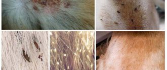
Maggots are easy to spot; they actively scurry around in the animal’s fur. The nasty-looking parasites devour the rotting flesh of the victim alive, thereby causing unbearable pain to the dog. Infection with maggots is accompanied by the following symptoms:
- apathy;
- aggression;
- severe itching;
- lethargic behavior;
- lack of appetite;
- intoxication of the body;
- heat.
Purulent wounds are a favorite place for maggots. They reproduce at maximum speed, since the dog’s body has all the comfortable conditions for their living: available food and warmth. In an open wound on a dog, you can easily notice elongated light-colored creatures with the naked eye. By feeding on the pet’s flesh, they will actively increase in size and absorb more and more soft tissue, which subsequently begins to decompose. Fly larvae, penetrating deep into wounds, eat through subcutaneous tunnels, causing even more harm to the dog. By eating away cells, maggots injure muscle and bone tissue. In addition, the spread of pathogenic microflora in the body further worsens. The skin tissue in the affected areas begins to swell and fester.
Transmission routes and carriers
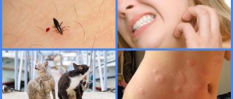
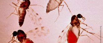
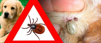
Since the larvae are transmitted to future carriers through the bites of the ubiquitous sand flies and mosquitoes , it can be extremely difficult to protect the dog from infection. The natural hosts of worms are foxes, raccoons, raccoon dogs and wolves. The carriers, as we have already said, are mosquitoes and mosquitoes, in whose bodies the larvae mature to the required stage. It is believed that infection in dogs leads to the development of the disease in 78% of cases. The remaining 22% are larvae for some reason, either die, or the pathology leads to the appearance of subcutaneous dirofilariasis in the pet.
For reasons still unknown, most of these cases are recorded in India (and not so much in animals, but in people). Perhaps the parasite has somehow changed on the Eurasian continent, or there are some other factors that prevent its “full” development in the lumens of large blood vessels and the cavities of the heart. In our country, cases of infection with “heart-subcutaneous” helminth are also becoming more common.
Parasitic diseases of dogs
Based on their habitat, parasites can be divided into four main groups:
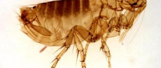
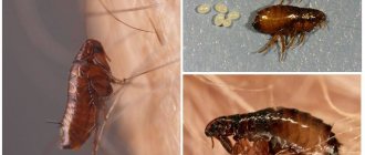
- external, living on the outer integument of the host (leeches, blood-sucking mosquitoes, ticks, fleas, lice eaters, etc.);
- skin parasites (itchy itch);
- cavity parasites that live in body cavities in contact with the external environment;
- internal - parasites of the blood, intestines and other organs (ascaris, trichinella, Plasmodium malaria, etc.).
A dog becomes infected with some parasitic diseases from other domestic animals (for example, a sheep is the host of 88 species of helminths, and a cow is the host of 55) and even from humans (so-called protozoal diseases).
Publications 1 - 6 of 6 Home |
Prev. | 1
| Track. | End Lice-eaters are small, up to 1-2 mm, wingless insects that infect the hair of animals. The disease is widespread. Dogs become infected from sick animals; stray dogs are especially dangerous in this regard.
-Go to publication-
Our pets are very often carriers of helminths or worms. Both adult dogs and small puppies become infected with worms. It is very important to detect worms in time and carry out their destruction. Some types of worms can be transmitted from a sick animal to humans, especially children. The dog must be dewormed regularly.
-Go to publication-
Demodicosis is a skin disease of many animal species caused by mites of the genus Demodex. Ticks are species specific, meaning that they do not move from one animal species to another. Normally, mites live on the skin in very small quantities and do not cause any problems to the animal, but when immunity decreases, they begin to multiply rapidly, leading to disease.
-Go to publication-
Dirofilariasis is a disease caused by helminths of the genus Dirofilariasis (Dirofilariasis from the Latin diro, filum - “evil thread”). The disease is common in the southern regions of our country, the countries of the Mediterranean, the Middle East, Africa and Asia, etc., but in recent years it has become frequently reported in Moscow and other regions of Russia.
-Go to publication-
Sarcoptic mange is a contagious, itchy skin disease of dogs (mange). Caused by mites of the Sarcoptes Canis species. This is an intradermal parasite.
-Go to publication-
Ehrlichiosis is a rickettsiosis and is transmitted by ticks. The pathogen is initially a tick-borne parasite, and therefore ticks form a reservoir of the pathogen. If you suspect ehrlichiosis, it is very important to contact your veterinarian promptly. If treatment is delayed, the prognosis may be unfavorable.
-Go to publication-
Publications 1 - 6 of 6 Home |
Prev. | 1 | Track. | End
Morphological features
It all starts with the appearance of small swellings and thickening on the surface of the skin. At first, their size does not exceed 1x1 cm, but within a few weeks the area of the lesions increases two to three times. Most often, these tumors are found on the lower surface of the abdominal wall, closer to the genital area. Soon the seals soften somewhat, becoming similar in consistency to cysts . When they were surgically removed, the inside of the cysts was separated from the “main” tissue by a demarcation line of inflammation (but not always). In addition, a worm is found inside the tumors. The length of subcutaneous parasites does not exceed 12 cm , their body is thin, grayish-white in color.
Development cycle
The adult worm lives in the subcutaneous tissues of the final host and... this is where the difficulties begin. Some hosts have microfilariae in their blood, some do not. What this is connected with is not completely known. Let us remember that these parasites are dioecious. The subcutaneous form of existence is not typical for them in nature. Hypothetically, worms should not and cannot reproduce under such conditions. There are several explanations for this phenomenon:
- Firstly, microfilariae can appear in the dog’s body after “additional” bites from insect carriers. The presence of adult parasites, as you might guess, is not required for this.
- Secondly, pulmonary aortas and heart may already have parasites. Who said that microfilariae must settle under the skin?
- The most exotic theory is that some parasitologists believe that under certain conditions, parasites of the genus Dirofilaria can “mutate” , turning into bisexuals. But this is very, very doubtful; this assumption has never been confirmed in laboratory conditions.
So, the development cycle of worms is closely related to mosquitoes and mosquitoes. In order for first-phase larvae to enter the insect’s body, it must suck blood from an infected host whose blood vessels already contain adult and actively reproducing parasites. The “mosquito” stage of development takes approximately two weeks from the larvae , during which they mature to the third stage. After this period, they are introduced into the salivary glands of the bloodsucker and wait in the wings. When a mosquito bites a dog, microfilariae of the third stage enter his bloodstream and begin to travel through the tissues and organs of the host. It is believed that at this stage of infection there are no clinical signs, but everything is somewhat different...
Preventive measures
Increased attention to the dog, regular inspection of the skin and timely treatment of open wounds with foul-smelling products will prevent flies from laying eggs. If at least one fly larva is noticed in the house, it is necessary to urgently inspect the animal and carry out a complete disinfection of the room. Regular control of flying insects, keeping the dog's habitat clean and maintaining hygiene will prevent the development of miasmas.
Maggots usually appear in a dog's wound if the animal is not properly cared for. Only absolutely irresponsible owners or a difficult street life can bring a pet to this state. A well-fed and healthy dog that has been regularly examined by a veterinarian can only in extremely rare cases be infected with parasites.
Clinical picture
So, what symptoms can indicate the presence of third stage larvae in a dog’s body? Firstly, a short-term increase in body temperature is possible. However, in 99% of cases this goes unnoticed. The dog may not look too good for a couple of days, his appetite is somewhat reduced , but that’s all. But blood tests often reveal eosinophilia, which is, in principle, characteristic of all types of helminthiasis . It is interesting, however, that during clinical studies an interesting fact was revealed: for some reason, an increase in the number of eosinophils is more typical for cases when a female Dirofilaria develops in the animal’s body. What this is connected with is not very clear. Most likely, the matter is in the cytological differences in the outer cuticle of female and male parasites. It is also interesting that in more than 70% of cases of subcutaneous dirofilariasis, when the cyst is dissected, it is female worms that are found . Again, why their survival rate in uncharacteristic conditions is so higher is a question that needs further study.
Therapeutic techniques
What treatment helps with subcutaneous dirofilariasis? Firstly, the worms themselves are removed surgically : the cysts are opened, the parasite is pulled out, the resulting cavity is washed with antiseptic and disinfectant solutions to avoid the development of inflammatory processes. If there are no other options, everything can be covered thickly with any veterinary powder based on streptocide . In general, there will be no special problems with larvae that have already penetrated under the skin. Owners with at least basic veterinary skills can handle this at home.
But it's not that simple. What to do with microfilariae currently in the animal’s blood? And they almost certainly exist, because if your pet is sick with dirofilariasis, then you probably live in an area unfavorable for this disease! Fortunately, there is nothing complicated here either: any anthelmintic drug that acts against parasitic nematodes will do. It is recommended to give the animal two courses of water, with an interval of ten days between taking the medicine. Please note that these drugs may not have an effect on larvae already under the skin. So in the case where microfilariae remain in the animal’s body, they may have to be removed in the future (using the surgical method described above).
But! I would like to remind you that subcutaneous helminths in dogs in this case are the least of the possible problems. Please note that adult heartworms prefer to live in the lumens of blood vessels and the cavities of the heart. So take the time to take your dog to the veterinarian for a full medical examination. It is quite possible that it will save not only the health, but also the life of the pet.
Treatment: how to remove maggots from a dog
Maggots on a dog simply need to be treated
.
How to remove maggots from a dog is a matter of life and death for the pet. On the Internet you can find many folk recipes for getting rid of this scourge, ranging from tar to simple chlorhexidine. However, you should pay attention to the health of your pet from the inside. Remember the last time deworming was carried out
.
Call a doctor at home? As soon as possible. Maggots in dogs
pose a significant danger to the animal.
- Maggots can be hatched
- First of all, you need to deworm the animal with appropriate medications, which will be recommended by your veterinarian.
- After deworming, you should thoroughly disinfect the room, or even better, the entire apartment in which the dog lives.
- All traditional methods of treatment should be used only after consultation with a veterinarian.
in several ways, but first you should carry out general preparation of the animal.
Although maggots are not contagious to humans
, but they have a fairly good appetite and
can eat an entire dog alive
.
If these maggots were noticed in the tail area, the open wound should be monitored for development of the situation. In the event that there are not many insects,
you can try to cope on your own, and
after treating the wound with chlorgesidine,
begin to destroy the uninvited guests. This is done as follows.
- How to remove maggots from a dog if there are not many of them:
- Apply liquid tar. Within a month, this substance will completely destroy maggots. The smell is not pleasant, but for the sake of the health of your beloved pet it is worth being patient with.
- When wondering what to do if your dog has maggots, you should remember that the use of bleach is strictly prohibited. This extremely toxic substance can lead to severe poisoning and death of the pet.
Nevertheless, maggots also have benefits; they remove dead areas in the wound. But this is noticeable only when these insects are specially placed in the wound, and the maggots themselves are the brainchild of a scientific laboratory.