Human brain cells
Thanks to MRI and EEG technology, it has become possible to take some incredible pictures of slices of the human brain. One of them was provided by Dr. Maria Prado-Figueroa. In the photograph, a cross-section of brain cells is magnified 63 times.
Rear-wheel drive Mazda SUVs coming in 2022 with Skyactiv engine
Fast food, white bread, alcohol: foods that harm female fertility
The husband dressed his pregnant wife all week: what outfits did she have to wear?
The tip of the butterfly tongue
No one will argue that butterflies are amazingly beautiful insects. Their multi-colored wings are full of different colors and patterns. But did you know that a butterfly’s tongue is just as colorful and beautiful? You can see this mesmerizing splendor only by magnifying their tongue 720 times.
This narrow house seems inconspicuous only from the outside, once you look inside
The famous Indian chef clearly showed how to boil perfect eggs: video
"Children need happy parents." Natalie Nevedrova named the reason for the divorce
Insects under a microscope: photos with names
materials » Microscopes » Articles about microscopes, microspecimens and studies of the microworld » Insects under a microscope: photos with titles
If you ever come across an enthusiastic microbiology enthusiast, sooner or later you will hear from him rave reviews about what he recently managed to observe. Bacteria, fungi, sponges, insects, parts of plants and animals - you will be bombarded with a huge amount of information and emotions. But hearing about it is not the same as seeing it for yourself. If you want to know what insects look like under a microscope, what onion skin looks like under 100x magnification, and what muscle tissue is made of, we will now tell and show you everything.
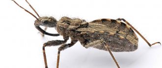
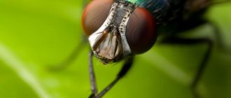
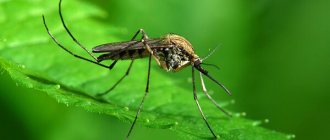
The most exciting thing about amateur microbiology is studying familiar objects at high magnification. Therefore, insects become one of the first objects for observation. Under a microscope, you can examine in detail the wings of bees and flies, study the villi on the legs of a fruit fly, and examine the structure of a mosquito's proboscis.
Insects under a microscope: photo gallery
| Bee wing, 60x | Drosophila, 60x | Mosquito mouthparts, 60x |
However, catching a nimble insect and placing it under a microscope lens can sometimes be difficult. Plants and mushrooms are more accessible for research. It is interesting to examine cereals, flower buds and mold under a microscope. At low magnification you can study the structure of plants, and at high magnification you can observe the division of their cells.
Plants and fungi under a microscope: photo gallery
| Onion peel, 150x | Mucor mold (dark field), 150x | Mitosis in onion root, 600x |
The next objects of microbiological research are usually animal and human tissues. To obtain them, it is enough to take a set of ready-made microslides. With its help, you can see nerve fibers, examine bone tissue, and study human blood. And it's much more exciting than insects under a microscope! Several photos with names can be seen below.
| Bone tissue, 150x | Mammal sperm, 600x | Loose connective tissue, 600x |
In this article we have presented only a small part of what can be seen under a microscope. And the actual experience gained cannot be compared in terms of the power of emotions with viewing photographs on the Internet. Studying the microworld is a fascinating hobby that is now available to many. Modern technologies make it possible not only to study tiny creatures, but also to photograph them yourself using a regular smartphone. Colorful photo albums of research can be obtained even with the help of an inexpensive amateur microscope.
4glaza.ru July 2017
Use of the material in its entirety for publicly accessible publication on media and any formats is prohibited. It is allowed to mention the article with an active link to the website www.4glaza.ru.
The manufacturer reserves the right to make any changes to the price, model range and technical specifications or to discontinue production of the product without prior notice.
| see also |
Other reviews and articles about microscopes, microslides and the microworld:
- Video! Levenhuk 870T microscope: video comparison of filtered and unfiltered water (MAD SCIENCE channel, Youtube.com)
- Video! Levenhuk 870T microscope: life in a drop of water from a swamp (MAD SCIENCE channel, Youtube.com)
- Video! Levenhuk 870T microscope: video of radioactive water (MAD SCIENCE channel, Youtube.com)
- Video! Levenhuk 870T microscope: video review (MAD SCIENCE channel, Youtube.com)
- Video! Levenhuk 870T microscope: video of salt water (MAD SCIENCE channel, Youtube.com)
- Levenhuk MED medical microscopes: review article on levenhuk.ru
- Video! Portable microscope Bresser National Geographic 20–40x and other children's devices in the line: video review (Tatyana Mikheeva channel, Youtube.com)
- Books of knowledge published by Levenhuk Press: detailed review on the website levenhuk.ru
- Video! Book of knowledge in 2 volumes. "Space. Microworld": video presentation (LevenhukOnline channel, Youtube.ru)
- Video! Video of bacteria under a Levenhuk Rainbow 2L PLUS microscope (channel “Microworld under a Microscope”, Youtube.ru)
- Levenhuk Rainbow 50L PLUS microscope review on levenhuk.ru
- Video! Detailed review of the Levenhuk LabZZ M101 series of microscopes for children (Kent Channel TV, Youtube.ru)
- Review of the Levenhuk LabZZ MTB3 set of optical equipment (microscope, telescope and binoculars) on levenhuk.ru
- Video! Levenhuk DTX 90 microscope: unboxing and video review of a digital microscope (Kent Channel TV, Youtube.ru)
- Video! Video presentation of a fascinating and colorful book for children “The Invisible World” (LevenhukOnline channel, Youtube.ru)
- Video! Great review of the Levenhuk 3S NG biological microscope (Kent Channel TV, Youtube.ru)
- Levenhuk Rainbow 2L PLUS microscopes
- Video! Levenhuk Rainbow and LabZZ microscopes (LevenhukOnline channel, Youtube.ru)
- Levenhuk Rainbow 2L PLUS Lime Microscope\Lime. Studying the microworld
- Choosing the best children's microscope
- Video! Levenhuk Rainbow 2L microscopes: video review of a series of microscopes (LevenhukOnline channel, Youtube.ru)
- Video! Levenhuk Rainbow 2L PLUS microscopes: video review of a series of microscopes (LevenhukOnline channel, Youtube.ru)
- Video! Levenhuk Rainbow 50L microscopes: video review of a series of microscopes (LevenhukOnline channel, Youtube.ru)
- Video! Levenhuk Rainbow 50L PLUS microscopes: video review of a series of microscopes (LevenhukOnline channel, Youtube.ru)
- Video! Levenhuk Rainbow D2L microscope: video review of a digital microscope (LevenhukOnline channel, Youtube.ru)
- Video! Levenhuk Rainbow D50L PLUS microscope: video review of a digital microscope (LevenhukOnline channel, Youtube.ru)
- Levenhuk Rainbow 50L Biological Microscope Review
- Video! Video review of Levenhuk Rainbow 2L and 2L PLUS school microscopes: the best gift for a child (KentChannelTV channel, Youtube.ru)
- Video! How to choose a microscope: video review for lovers of the microworld (LevenhukOnline channel, Youtube.ru)
- Photo gallery! Sets of ready-made microslides Levenhuk
- Microscopy: dark field method
- Video! “One day of a slipper ciliate”: video filmed using a Levenhuk 2L NG microscope and a Levenhuk digital camera (LevenhukOnline channel, Youtube.ru)
- Video! Review of the Levenhuk Rainbow 2L NG Azure microscope on the Karusel TV channel (LevenhukOnline channel, Youtube.ru)
- Levenhuk Fiksiki Fire Microscope Review
- Compatibility of Levenhuk microscopes with Levenhuk digital cameras
- How does a microscope work?
- How to set up a microscope
- How to care for a microscope
- Types of microscopes
- Technique for preparing microslides
- Photo gallery! What you can see with Levenhuk Rainbow 50L, 50L PLUS, D50L PLUS microscopes
- Grid or scale. Microscope and the ability to take precise measurements
- Ordinary objects under a microscope lens
- Insects under a microscope: photos with names
- Ciliates under a microscope
- Invention of the microscope
- How to choose a microscope
- What do leukocytes look like under a microscope?
- What is a laser scanning microscope?
- Fluorescent microscope: the price is high, but justified
- Microscope for soldering microcircuits
- Immersion microscope system
- Measuring microscope
- Microscopes from the largest professional models to simple children's ones
- Professional digital microscope
- Force microscope: for serious research and fun
- Dental treatment under a microscope
- Human blood under a microscope
- Halogen lamps for microscopes
- French experiments - microscopes and development kits from Bondibon
- Sets of preparations for a microscope
- Adjusting the microscope
- Microscope for electronics repair
- Operating microscope: price, capabilities, applications
- “Scale microscope” - what optical device is called that?
- Wart under a microscope
- Viruses under a microscope
- Operating principle of a dark-field microscope
- Cover glasses for microscopes – should I buy them or not?
- Optical microscope magnification
- Optical design of the microscope
- Diagram of a transmission electron microscope
- The device of an optical microscope at a theodolite
- Fungus under a microscope: photos and research features
- Why do you need a digital camera for a microscope?
- Microscope stage - what is it and why is it needed?
- Transmitted light microscopes
- Organelles discovered using an electron microscope
- Spider under a microscope: photos and study features
- What does a microscope consist of?
- What does hair look like under a microscope?
- Eye under a microscope: photos of insects
- DIY microscope from a webcam
- Bright field microscopes
- Mechanical microscope system
- Microscope lens and eyepiece
- USB microscope for computer
- Universal microscope - does such a thing exist?
- Sand under a microscope
- Ant through a microscope: studying and photographing
- Plant cell under a light microscope
- Digital industrial microscope
- Human DNA under a microscope
- How to make a microscope at home
- The first microscopes
- Stereo microscope: buy or not?
- What does a cancer cell look like under a microscope?
- Metallographic microscope: should I buy it or not?
- Fluorescence microscope: price and features
- What is an "ion microscope"?
- Dirt under a microscope
- What does a tick look like under a microscope?
- What does a worm look like under a microscope?
- What does yeast look like under a microscope?
- What can you see with a microscope?
- Why are research microscopes needed?
- Bacteria under a microscope: photos and observation features
- What does the microscope lens aperture affect?
- Roundworms under a microscope: photos and study features
- How to use slides for a microscope
- We study GOST: microscopes that meet standards
- Instrumental microscope – buy or not?
- Where to buy a reading microscope and why is it needed?
- Atom under an electron microscope
- How a mosquito bites under a microscope
- What does a fly look like under a microscope?
- Amoeba: photo under a microscope
- A savvy flea under a microscope
- Lice under a microscope
- Bread mold under a microscope
- Teeth under a microscope: photos and observation features
- Snowflake under a microscope
- Butterfly under a microscope: photos and observational features
- The most powerful microscope – how to choose the right one?
- Leech mouth under a microscope
- Midge under a microscope: jaws and body structure
- Microbes on hands under a microscope - how to see?
- Water under a microscope
- What does a worm look like under a microscope?
- Cell under light microscope
- Onion cell under a microscope
- Brains under a microscope
- Human skin under a microscope
- Crystals under a microscope
- The main advantage of light microscopy over electron microscopy is
- Confocal fluorescence microscopy
- Probe microscope
- Operating principle of a scanning probe microscope
- Why is it difficult to make an X-ray microscope?
- Macroscrew and microscrew of a microscope - what are they?
- What is a tube in a microscope?
- Main plane of the polarizer
- What is affected by the angle between the main planes of the polarizer and analyzer?
- Purpose of the polarizer and analyzer
- Study method - microscopy in practice
- Microscopy of urine sediment: interpretation
- Analysis "Smear microscopy"
- Scanning electron microscopy
- Light microscopy methods
- Optical microscopy (light)
- Light, fluorescence, electron microscopy - different research methods
- Dark-field microscopy
- Phase contrast microscopy
- Natural light polarizers
- Scottish physicist who invented the polarizer
- Focusing mechanism in a microscope
- What is a field diaphragm?
- Microscope Micromed: instruction manual
- Mikmed microscope: instruction manual
- Where can I find the instructions for the LOMO microscope?
- Micros microscopes: user manual
- What is the function of the clamps on a microscope?
- Microscope lens working distance
- Do-it-yourself microscope for a microscope
- Hanging drop method
- Crushed drop method
- Tardigrade under a microscope
- Golgi apparatus under a microscope
- What to do with children at home?
- What to do during quarantine at home?
- What should schoolchildren do during quarantine?
- Choosing a microscope: do reviews matter?
- Microscope for a schoolchild: which one to choose?
- A little about the wholesale purchase of microscopes and other optical equipment
- How much does a magnifying glass magnify?
- Where to buy a magnifying lamp - a cosmetology model with backlight?
- Which magnifying lamp to buy for manicure?
- Is it possible to buy a magnifying lamp for eyelash extensions in an online store?
- Cosmetology magnifying lamp on a tripod: should I buy it for home or not?
- Binocular magnifier with accessories
- What does a numismatist's magnifying glass look like?
- Magnifier lamp – handicraft magnifying glass with backlight
- “Magnifying glass on a stand” - what kind of optical device is this?
- Magnifying glass – projector for enlarged images
- Making a magnifying glass with your own hands
- Basic functions of a magnifying glass
- Where can I find a magnifying glass?
- Binocular magnifying glass – the price of opportunity
- Stationery magnifying glass: choosing optical equipment for the office
- What does coronavirus look like under a microscope?
- What is the name of the main part of the microscope?
- Where to buy power supplies for a microscope?
- Microscope lens structure
- What foods look like under a microscope
- What will the microminiature museum show?
- Features and application of cell staining methods
Ant head
Watching the hard-working ants, we see how they build their homes or try to steal crumbs of food from our table. But if you see a cute goosebump at tenfold magnification, this hard worker will not seem like a harmless bug.
By enlarging familiar insects and plants, you can understand how complex and unique the world around us is. And also how fragile it is, how easily we humans can destroy the beauty that surrounds us.
Found a violation? Report content
Danger of cockroaches
These insects are dangerous, first of all, because they are carriers of infectious diseases and worms. As they crawl over our food and utensils, they leave behind millions of different microbes. If you do not treat such dishes, you can become infected with something very unpleasant. This is due to the fact that this insect can eat not only food debris or garbage, but also the feces of various animals and even human waste.
If you dreamed that you were bitten by a cockroach, then it may not be a dream. These insects can, in fact, bite humans. This occurs as a result of lack of food or water. In such cases, the barbel does not show any special aggression. Simply, in this way, it makes up for the lack of food. But this is very dangerous, as the bites can become inflamed and fester. In addition, they have the custom, when children are sleeping, of biting their eyelashes and eyebrows, as well as gnawing the epidermis in the corners of their lips.
Numerous cases of allergic reactions after bites also confirm the danger of these insects. So if you dreamed of cockroaches and their antennae, then it is better to check your home and treat all suspicious places.


