Representatives of the family of flies - gadflies - have a hemispherical, well-developed head, with bare eyes, which in females are spread wider at the back of the head than in males; There are three simple eyes. The antennae are placed in a pit on the forehead, short, segmented, with bare or half-pinnate bristles; the female has a significantly larger segment 3 than the male.
The proboscis of the American group Cuterebridae is quite large, horny, geniculate, retracted into the mouth slit and is hardly noticeable from the outside, without tentacles. The body is large, wide, with a transverse seam on the back, the hind legs are often very elongated.
Dogs become infected when they come into contact with grass in nature that contains botfly larvae. The movement of the dog in relation to the grass stimulates the larva to move towards a moving object - towards the dog. The larva will then move around the dog's body until it finds an opening under the skin.
Infection with gadfly larvae is seasonal from summer to early autumn, during the period of activity of adult gadflies.
An infestation of Cuterebra botfly larvae may present as bumps above the surface of the skin, or the dog may show signs associated with the larvae moving through the tissue. Symptoms may include respiratory signs, neurological symptoms, ophthalmological (eye) and skin symptoms.
Respiratory symptoms:
- Cough.
- Fever.
- Hesitant breathing.
Neurological symptoms:
- Dizziness.
- Loss of coordination (movement in a circle).
- Paralysis.
- Blindness.
- Constant “lying down” position.
Ophthalmology symptoms:
- Damage caused by larvae in the eyeball.
Skin symptoms:
- The location of the larva under the skin (tubercle, lump, compaction) will be raised above the skin level and has a hole so that the larva can breathe.
Treatment of botfly larvae infestation in dogs
If the larva is at the end of its migratory stage and has settled in a specific location on the body, such as under the skin, eye or nose, your veterinarian will be able to remove it safely.
Myiasis is a collective concept rather than the name of a specific disease. Unfortunately, not all animals, even those living on private property, are subject to daily inspection, scratching, and bathing. Meanwhile, they may have wounds, abrasions, violations of the integrity of the skin, and all this goes unnoticed by the owner. The open wound surface attracts insects with the smell of blood and flesh, and sometimes pus. So the flies strive to lay their larvae there so that their offspring have something to eat.
The main culprit of myiasis is the Wohlfarth fly, and the disease is called “Wolfartiosis” (blackening of wounds).
Main routes of penetration into animals and humans
How exactly the gadfly lays eggs on its prey and where the larvae will develop depends on what type of insect it is. There are three main groups of gadflies:
- subcutaneous - the female lays eggs on the fur or on the skin, the hatched larvae penetrate inside and travel through the tissues;
- gastric - the female lays eggs on food plants, the larvae enter the animal’s digestive system with food and feed on the tissues of the internal organs;
- cavitary - a viviparous female in flight injects larvae into the animal’s face, the parasites penetrate into the eyes or nose, and through them further to the brain.
The only species of gadflies that attack humans are those whose larvae develop under the skin. But people can become infected with absolutely any type of parasite. The female may throw the larvae in the face, or you can eat meat infected with the larvae of the gastric botfly. Even petting an infected cow or sheep can produce one or more larvae.
Another way in which gadfly larvae appear in the human body is through blood-sucking insects. The female lays eggs on the abdomen of the mosquito, which lands on a person to bite him, and the egg ends up on the skin. Body heat provokes the appearance of a larva that penetrates the skin.
Description of the pathogenic insect
The insect itself feeds on the juices of the plant. 12-15 days after mating, the female lays larvae. To do this, she looks for living tissue, wound surfaces, and macerated (wrinkled, inflamed) animal skin. It lays 10-20 larvae in wound openings and folds of skin. From this moment myiasis begins. The larvae develop quickly; in a week they are able to molt twice and grow up to two centimeters.
Mature larvae fall out of the wound and burrow into the ground, where they pupate. If the environment is suitable, the ground is warm and soft, then complete pupation occurs in 10-12 days, otherwise it is delayed or suspended. If the larva falls out in late autumn, then it overwinters in the ground, in the pupal phase, and continues its development in the spring.
Wohlfarth fly lays larvae mainly on animals
The fly itself is a two-winged insect from the blowfly family. Very common in southern Russia, the Middle East, and China.
Wolfarthiosis mainly affects sheep farms. However, other animals, dogs, cats, and sometimes humans, can also develop myiasis.
Course of the disease
In cats and dogs, the disease is rarely recorded due to the physiological characteristics of these animals. Licking your own wounds is the best way to promote both wound hygiene and healing. However, in hard-to-reach places, where the cat cannot reach and lick, long-term non-healing processes are possible.
Initially, myiasis does not cause much trouble. However, over time, when the wound surface does not granulate, it is necessary to urgently seek help from a veterinary specialist.
The larvae, penetrating deep into the wound of a cat or dog, are able to make subcutaneous tunnels.
Myiases are diseases of dogs and cats caused by the larvae of certain types of flies.
By eating away cells, they significantly damage connective and muscle tissue. In addition, myiasis is further aggravated by the spread of pathogenic microflora. The wound tissue begins to become inflamed (swell, fester).
Is it worth calling a doctor to your home to get help on how to remove maggots from a dog?
A doctor's visit to your home will help save a lot of time, which can be spent usefully, for example, examining the animal's rear hole for cleanliness. You should also carefully prepare for the doctor’s visit. Prepare a clean towel, wet wipes, and a large, wide surface covered with a sterile sheet. When the doctor arrives, you should remain calm and not panic. Dogs are excellent psychologists and they detect the slightest change in their owner’s mood. Panic is also easily transmitted to them, and an agitated animal is quite difficult to examine. Which doctor should I call? First of all, you should invite a veterinarian who will conduct a general examination of the animal. By agreement with the owner, a dermatologist from our veterinary center will be sent.
- You can further familiarize yourself with the list of our doctors who are ready to help on the first call:
If you use such a convenient service as a veterinarian at home at least once, you will no longer want to come to the veterinary center on your own.
Symptoms
Cutaneous myiasis in its acute form is manifested by anxiety in the animal. Superficial wounds are easily identified and treatment is carried out quickly and effectively.
If the myiasis is subcutaneous, with many “pockets,” then the animal is depressed or agitated, and the cat can become aggressive. Festering wounds cause itching, the animal tries to comb and scratch the affected area. If myiasis has affected deep-lying muscle tissue, and the process has become chronic, an increase in body temperature and severe depression of the animal is observed.
Treatment
When treating animals, you cannot get by with simply treating the wound surface. Until all the larvae are removed, the “pockets” of the wound are excised, the exudate is removed and the pathogenic microflora is suppressed, the process of tissue granulation will not begin.
Surgery and wound treatment can only be carried out by a qualified specialist, especially if muscle groups are affected. Treatment of wounds in cats should be performed under local anesthesia.
After cleaning the wound (mechanical removal of its contents), medical treatment is carried out, irrigating the cavity with antibiotic solutions (erythromycin or penicillin). Drainage is inserted into the treated “pockets”. Vishnevsky ointment is applied to the wound surfaces. In case of severe inflammatory processes, a course of treatment with antibiotics is prescribed.
Treatment consists of surgical removal of the larvae from the affected tissue
When treating small animals, cats, dogs, a fixing bandage must be applied after the manipulations.
To treat wounds without removing the larvae, the drug "Volfazol" is used. It contains chlorophos (FOS-organic pesticide - to destroy larvae), erythromycin (antibiotic - to suppress microflora), birch tar - a natural substance that enhances tissue keratoplasty. Release form: aerosol foam. The drug is easy to use and effective. To carry out treatment, foam is applied inside the wound and on the entire outer wound surface. The waterproof film formed after application protects the wound from wetting, infection and additional invasion.
"Wolfazol" is used for the prevention and treatment of all farm animals, as well as cats, dogs and other carnivores.
Preventive actions
Attentive attitude towards animals, timely examination of the skin and veterinary measures will help prevent the development of myiasis.
Timely treatment of wound surfaces with iodoform and aerosol foul-smelling substances will not allow insects to lay their larvae in the wound.
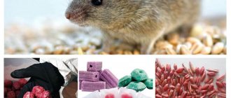
Regular control of insects - laying out poisonous baits, hanging Velcro, keeping places where people and animals live clean - will significantly reduce the spread and reproduction of dipterans.
Insects accompany humans and animals everywhere, however, humankind can control their numbers. Modern agrochemistry has created an incredible number of means to combat flies, mosquitoes, and ticks; you just need to use them in time.
It is a disease caused by the fly Wohlfahrtia magnifica.
.
It is one of the varieties of Myiases, that is, diseases caused by the development in the tissues of fly larvae that feed on dead and living tissues of the host. There are dozens of similar pathologies in the world, but in our area, wolfarthiosis is the most common
.
The disease was first described back in 1862. It is widespread throughout Central and Southern Europe, Russia, Central Asia, and China. The disease is particularly common in Italy and Hungary, and multiple cases have been reported in Morocco.
Female flies Wohlfahrtia magnifica have an excellent sense of smell and are able to find potential hosts by smell from many tens of kilometers away. It is worth noting that the insect never attacks completely healthy dogs - there must be signs of inflammation on the animal’s skin. Flies react especially
Development cycle
The female flies up to the future “incubator” and literally injects the larvae into the affected area of the skin.
Each “injection” contains at least 10-15 individuals. The larvae quickly burrow into the wound, actively feed and molt twice. After about five days (depending on the ambient temperature), they crawl out of their cozy nest, fall onto the soil, burrow into it, and pupate. After one to three weeks, the imago (that is, the adult stage) emerges.
Interesting!
Adult flies are most active at an ambient temperature of 20-27 degrees Celsius. If it gets hotter, they find shade and fall into a kind of torpor.
Symptoms and treatment methods
What are the symptoms? Determining the presence of the disease is simple - even an inexperienced breeder can easily see fatty larvae freely crawling in an inflamed, smelly wound, from which large volumes of pus are released. The surface of the wound lesion sometimes reaches the area of the palm of an adult; it is covered with brown, watery contents with a disgusting odor.
In severe cases, the animal’s condition quickly deteriorates due to the large volume of toxins
. When the dog licks the wound surface, everything becomes even worse, since the wound is further macerated (softened), and even more pathogenic microflora penetrates there in addition to the larvae.
As a rule, maggots appear in a dog’s wound if the animal is neglected. Untreated open injuries, festering wounds, ulcers, scratched insect bites are an ideal environment for laying eggs of various flies, from which larvae later hatch. This phenomenon in clinical conditions is called miasm. If the necessary measures are not taken urgently, this can lead to serious consequences, including the death of the pet.
Treatment technique
To get the right maggots, the flies are kept in a sterile, enclosed area where they can lay their larvae. The worms are then placed in bags and only then can the “live medicine” be used. The effects of worms on the human body are as follows:
- sterilization of focal wounds;
- stimulation of healing;
- cleansing by eating necrotic areas;
- the secreted substance allantoin stimulates the healing process.
Fact! Allantoin, secreted from the urea of the larvae, is also found in cow urine. That is why in villages they still wash wounds with evaporated cattle urine.
It should be noted that seraticin, an antibiotic contained in the mucus of worms, resists 12 strains of methicillin-resistant Staphylococcus aureus, destroys E. coli and bacteria that cause pseudomembrane colitis.
Larval therapy includes several mandatory stages:
- breeding larvae of a certain type of fly (green fly, blowfly);
- obtaining eggs with their subsequent washing and sterilization;
- hatching larvae;
- placing worms in the wound;
- opening the wound and removing maggots.
Before placing worms into a wound, they are forced to starve, so placing larvae in a wound for more than a day is not recommended. However, sometimes the time of therapeutic effect is calculated individually. It all depends on the severity, size of the affected area, type of wound and the presence of purulent inflammation. For example, if the lesion is chronic, the bed is covered with disinfected larvae for 4 days. The procedure can be repeated several times, achieving complete cleaning of the wound bed and a speedy recovery of the patient.
Interesting! Sometimes silky lucilia larvae are used. These worms secrete an enzyme that dissolves dead tissue and then eat the resulting substance. After 2-4 days, individuals grow to a size of 12 mm and stop cleaning the wound. If necessary, they are replaced with a new portion of maggots and therapy is continued.
Unfortunately, larval therapy in its original form causes more rejection than acceptance in the average patient. Not every person can allow worms to appear in a wound, and doctors prefer more conservative methods of healing. But if the doctor suggests trying this option, you should not refuse - in just 1-2 days the bed of the most advanced wound will be cleared, and the healing process will go much faster. In this case, you will not have to administer loading doses of antibiotics and other anti-inflammatory medications.
As a rule, maggots appear in a dog’s wound if the animal is neglected. Untreated open injuries, festering wounds, ulcers, scratched insect bites are an ideal environment for laying eggs of various flies, from which larvae later hatch. This phenomenon in clinical conditions is called miasm. If the necessary measures are not taken urgently, this can lead to serious consequences, including the death of the pet.
How to detect maggots?
Purulent wounds are a favorite place for maggots. They reproduce at maximum speed, since the dog’s body has all the comfortable conditions for their living: available food and warmth. In an open wound on a dog, you can easily notice elongated light-colored creatures with the naked eye. By feeding on the pet’s flesh, they will actively increase in size and absorb more and more soft tissue, which subsequently begins to decompose. Fly larvae, penetrating deep into wounds, eat through subcutaneous tunnels, causing even more harm to the dog. By eating away cells, maggots injure muscle and bone tissue. In addition, the spread of pathogenic microflora in the body further worsens. The skin tissue in the affected areas begins to swell and fester.
How does the larva develop?
Reproduction and reproduction of offspring is the main purpose of the existence of an adult gadfly. As soon as she is born, the female immediately goes in search of a male to mate with. After fertilization, the female looks for a suitable object on which to lay eggs.
The female subcutaneous gadfly lays eggs on the skin of the animal, the gastric one on the food plant, the cavitary one is viviparous, so it immediately attaches larvae to the victim. Once inside the body of a person or animal, the larva begins to actively eat. Her diet includes blood and tissues, from which she receives all the nutrients necessary for development and growth.
The larval stage lasts from 5 to 10 weeks. During this time, the parasite goes through several molts and increases in size by 10–15 times. A fully formed larva is ready to become a pupa. To do this, it leaves the host's body. The larva may gnaw through the skin to escape or be shed in the feces.
Symptoms and treatment
Miasm in dogs is divided into three types:
If maggots have appeared in a dog’s wound, you cannot do only superficial treatment. Treatment should be carried out under the full supervision of a veterinarian. Since all the larvae are not destroyed, the exudate is eliminated and the pathogenic microflora is not suppressed, the tissues will not heal, which means that an outbreak of another lesion is possible.
Fly larvae are not dangerous to humans, but they have a good appetite and can eat a dog alive.
If the presence of maggots was discovered at the initial stage of reproduction and there are few of them, you can cope on your own. For treatment you will need chlorhesidine; it must be applied to the wound for several days. But it should be remembered that the solution contains toxic substances that the animal can lick off, and this can lead not only to severe poisoning, but also to the death of the pet. In this situation, it is better not to take risks and if maggots appear in the dog’s wound, immediately contact a veterinary clinic.
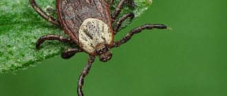
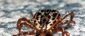
How to tell if your dog has skin parasites
Most parasites are difficult to notice, but their waste products speak for themselves. Although almost every owner of a four-legged friend knows what fleas look like. The easiest way to tell that a dog is infested with fleas is by its behavior. The animal itches furiously and bites off fleas.
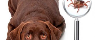
Mites are not so active (with the exception of scabies), but they are also easy to detect. It is enough to palpate your pet’s skin and inspect all suspicious bumps and bulges. In addition, it is quite possible that your pet will help find the parasite by scratching the bite site.
Now regarding the scabies mite and other skin parasites, whose activity does not manifest itself at first. I think you have already understood that contacting a veterinarian in such cases is the only opportunity to find out the root cause of the disease and nip the development of parasites in the dog in the bud.
Preventive measures
Increased attention to the dog, regular inspection of the skin and timely treatment of open wounds with foul-smelling products will prevent flies from laying eggs. If at least one fly larva is noticed in the house, it is necessary to urgently inspect the animal and carry out a complete disinfection of the room. Regular control of flying insects, keeping the dog's habitat clean and maintaining hygiene will prevent the development of miasmas.
Question for experts: how to remove maggots from a dog? There are so many of them and she suffers a lot. The dog is old and lives in a kennel in the yard
Best regards, Valentina Machneva
How do signs of maggots appear in dogs?
It is not difficult to notice maggots on a dog’s body - you just need to unroll the fur in certain places on the animal’s body. Sometimes, due to the thick fur, it is not possible to detect parasites right away, but the pet itself usually helps. Crawling, the larvae cause him a lot of anxiety, so a non-paralyzed dog will itch or try to lick the wound. Accordingly, the owner should pay increased attention to exactly the same area as the pet itself.
In advanced cases, deep holes appear on the body, formed by the larvae eating the flesh of a living creature. Taking a closer look at them, you will also probably notice worms, about 2-5 mm long.
Additional symptoms
By eating living flesh, fly larvae cause considerable discomfort to your pet, so it is not surprising that an infected dog becomes apathetic, lethargic and refuses food. Often, due to intoxication of the body, body temperature rises. If the pet does not refuse food, diarrhea may develop, although this symptom is quite rare.
Best answers
Nadezhda Mikhailova: horror! Why did you bring the dog to this? She's rotting alive!!!
This is me: she has already died, judging by the fact that maggots have appeared - about two weeks ago
Fox: Why are there maggots on her, are there wounds?
Irina Kozyavkina: well, you are sadists. I'll call the police right now
Elena Rusakova (Averina): you need to treat the wound with an antiseptic solution. Wash the maggots thoroughly with water. then put on a bandage with miramistin. In general, you can buy chlorhexidine and wash the wound every day. It is advisable to show the dog to the veterinarian later. or at least give a course of antibiotics, such as gentamicin. it is inexpensive, 2 times a day, 0.3 each. Don’t be afraid, prick in the leg, you will feel the muscle there.
kennel “Bas-ko-chi”: What a nightmare, imagine yourself in his place, here is another example of a dog “for yourself”, or for free, why care for it? Fiends
Marina Frolova: well, firstly, they are simply removed with peroxide or chlorhexidine with a cotton swab, secondly, this is how to run a dog so that maggots will appear, 3rd, cut off all the hair around the affected areas so that it does not get into the wounds . Apply a thin layer of Levomikol ointment, do not lick it off. Do this 2-3 times a day until complete recovery.
DIMON FILIN: she urgently needs to be taken to the veterinarian. Maggots only breed in rotting meat. and the process of rotting is underway. don't torture the dog, take it to the vet. clinic!
OLGA GALUK: how to cure maggots from a dog?
Angelika Byankina: This is to the vet!!! They need to be removed, not lotions applied!!!