Life cycle of development of malarial plasmodium in mosquito organs
When sucking the blood of a person with malaria, malarial plasmodia, which are at different stages of development, enter the mosquito’s body, but only gamonts (immature sexual forms) undergo further development. All other plasmodia die. In the stomach of a mosquito, malarial plasmodia travel a complex path.
Rice. 6. The development cycle of malarial plasmodium in the body of a mosquito. Female gamete (17). Formation of the male gamete (18). Fertilization (19). Ookineta (21). Oocyst development (22 and 23). Release of sporozoites from the oocyst (24). Sporozoites in the salivary gland of a malaria mosquito (25).
Maturation of germ cells
In the mosquito's midgut (stomach), gametocytes (immature sexual forms) are converted into gametes (mature sexual forms). Macrogametes or females are formed (mature) from macrogametocytes. Males are formed from microgametocytes. Moreover, from each microgametocyte up to 8 mobile snake-like microgametes are formed. It has been proven that if there are less than 1 - 2 gametocytes in 1 mm3 of the blood of an infected person, mosquito infection does not occur.
Rice. 7. In the stomach of a mosquito, male gamonts “throw out” flagella. The process is called exflagellation.
Fertilization
In 20 minutes. (up to 2 hours) fertilization occurs in the stomach of the mosquito: the microgamete is introduced into the female individual - the macrogamete. When the gametes fuse, they form a zygote. The body of the zygote elongates and it turns into a mobile ookinete. The nuclei of germ cells fuse.
Sporogony
Next, the ookinete penetrates the wall of the mosquito’s stomach, rounds and penetrates its outer wall, becomes covered with a protective membrane, grows and turns into an oocyst. The number of oocysts can be from a few to 500. The entire process from a mosquito bite to the formation of an oocyst lasts about 2 days.
Inside the oocyst, vigorous division of plasmodium nuclei occurs, around which areas of protoplasm condense. The nucleus with a section of protoplasm is called a sporoblast. Inside the sporoblasts, spindle-shaped sporozoites develop, the number of which can reach 10 thousand. The oocyst swells to such a size that the sporozoites float freely in it. The oocysts deposit a pigment, the pattern of which can be used to determine the type of plasmodium.
Rice. 8. The ookinete attaches to the inner wall of the midgut (photo on the left), penetrates it, becomes rounded and penetrates the outer wall, becomes covered with a protective membrane, grows and turns into an oocyst (photo on the right).
Rice. 9. A large number of oocysts on the outer lining of the stomach (a). Opened oocyst and many sporozoites (b). The photo on the right shows oocysts on the outer lining of the stomach.
After the oocyst shell ruptures, the sporozoites enter the body cavity and hemolymph of the mosquito and are spread throughout the body. The largest number of them (hundreds of thousands) accumulates in the salivary glands.
Rice. 10. The photo shows a section of the body of an infected Anopheles mosquito. A huge number of spindle-shaped sporozoites are visible in the hemolymph.
Rice. 11. In the picture on the left there are many sporozoites in the salivary gland of a mosquito. The photo on the right is a view of sporozoites.
After 2 weeks, sporozoites acquire virulence, maintaining infectious properties for up to 2 months. The sporozoites then degenerate.
The timing of sporogony is influenced by the type of mosquito and the ambient temperature.
When mosquitoes are infected with Plasmodium vivax, the insect becomes dangerous after 7 days, Plasmodium falciparum - after 8 - 10 days, Plasmodium malariae - after 30 - 35 days, Plasmodium ovale - after 16 days.
Dangerous symptoms
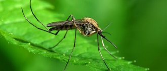
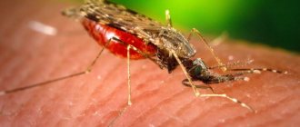
The genus Malaria mosquitoes has more than 400 species. Do not think that their representatives live only in African countries. They are distributed everywhere except in the northern regions. Characteristic features of adult insects are an elongated body, long legs and proboscis, and a short head. Their wings are covered with scales along the veins.
The bite of such a mosquito is the most common route of infection with malaria. But unsterile medical instruments can also be the cause. The first symptom of the disease is the appearance of fever. As red blood cells are destroyed, an increase in the size of the spleen, hardening of the liver, and the development of anemia are observed.
So, the life cycle of malarial plasmodium is characterized by a change of hosts: intermediate and final. The first is man. In his blood cells, asexual reproduction of plasmodium occurs through schizogony. The definitive host of the parasite is the mosquito. In his body, the parasite cells reproduce sexually. The development of malaria occurs only if it is possible to repeat the stages of the life cycle. Otherwise, a dangerous disease does not develop.
Life cycle of Plasmodium falciparum in humans: exoerythrocytic (preclinical) stage of malaria
Infection
When an infected female mosquito bites a person, malarial plasmodia at the sporozoite stage enter the human bloodstream with the saliva of the insect. Within 10 to 30 minutes, sporozoites move freely in the blood plasma and then settle in the liver cells. Some of the sporozoites (bradysporozoites) Plasmodium ovale and Plasmodium vivax hibernate, while another part of them, as well as Plasmodium falciparum and Plasmodium malariae (tachysporozoites) begin hepatic schizogony immediately.
Rice. 12. Tissue exoerythrocytic schizogony. 2 - trophozoite, 3 - schizont, 4 - release of merozoites from liver cells into the blood.
Period of tissue schizogony
In liver cells (hepatocytes), the transformation of sporozoites into tissue schizonts occurs, which after 6–15 days divide to form many tissue merozoites. From one sporozoite, from 10 to 50 thousand hepatic merozoites (schizonts) are formed, which enter the blood after 1 - 6 weeks.
When infected liver cells are destroyed, tissue merozoites are released into the blood. This ends the incubation period of malaria and begins the period of erythrocyte schizogony - the period of clinical manifestations.
Hibernation process
Some sporozoites (hypnozoites) of Plasmodium ovale and Plasmodium vivax, once in hepatocytes, turn into inactive forms and hibernate. Parasites can remain in this state for months and years, causing distant relapses.
Rice. 13. Tissue schizont in the liver.
Treatment of malaria
The main goal of therapy for this disease is to prevent the occurrence/recurrence of attacks and completely destroy the pathogen. The disease malaria or swamp fever is more common in endemic areas, so travelers should take preventative measures in advance. Treatment of malaria is carried out with the help of drug therapy, as a rule, Primaquine, Chloroquine, Atabrine (quinacrine hydrochloride), Akriquine are used.
Medicines for malaria
Drug therapy for this disease is considered an effective method. There are proven medications for malaria that have been used for a long time. An example of such a medication is Quinine, which was replaced by Chloroquine for a while, but then began to be actively used again. The reason for this was the emergence and then spread in Asia and Africa of Plasmodium falciparum, which was resistant to Chloroquine.
Depending on the region where the infection occurred, certain drugs against Plasmodium falciparum can be used. Most of them are suitable for both treatment and prevention. Artemisia annua extract containing artemisinin and analogues of synthetic origin are highly effective, but also high in cost. The disease poses a great danger to residents who live in endemic areas where there is no access to drugs. In developed countries, there are no problems with purchasing medicines.
- Eco-leather - what kind of material is it from the photo. Properties and characteristics of eco-leather, how to distinguish it from genuine leather
- Herpes in the nose: symptoms and treatment of the disease
- Effective exercises for the inner thigh
Life cycle of Plasmodium falciparum in humans: erythrocyte (clinical) stage of malaria
After the liver cells rupture, merozoites enter the blood and invade red blood cells. The erythrocyte (clinical) stage of schizogony begins.
Rice. 14. Erythrocyte schizogony. 5 and 6 are ring-shaped trophozoites. 7, 8 and 9 - young, immature and mature schizonts. 10 - erythrocyte merozoites.
Attachment to red blood cells
Attachment of merozoites to the membrane of erythrocytes and invagination into their membranes occurs due to the presence of special receptors on the surface of red blood cells. It is believed that the receptors on the surface of erythrocytes that serve as targets for merozoites are different for different types of Plasmodium.
Rice. 15. Red blood cells infected with Plasmodium vivax (the causative agent of tertian malaria) and Plasmodium ovale (the causative agent of tertian malaria) become enlarged, discolored and deformed, and toxic granularity appears in them. When infected with Plasmodium malariae (the causative agents of four-day malaria) and Plasmodium falciparum (the causative agents of tropical malaria), the shape and size of red blood cells do not change.
Erythrocyte schizogony
Having penetrated red blood cells, schizonts absorb the protein globin (a component of hemoglobin), grow and multiply.
In the erythrocyte, the parasite goes through 4 stages of development:
- Ring (trophozoid) stage.
- Stage of amoeboid schizont.
- Morula (fragmentation) stage. At this stage, the nuclei of schizonts are repeatedly divided (into 6 - 25 parts), and areas of the cytoplasm are separated around them. Erythrocyte merozoites are formed.
- Some parasites go through the stage of gametocyte formation.
A parasite that has penetrated an erythrocyte is called a trophozoid (schizont). It gradually increases in size. A vacuole appears near its nucleus and the trophozoid takes the form of a ring (ring) - a ring-shaped schizont.
As they grow (schizonts feed on hemoglobin), they increase in size and take on the appearance of an amoeba - an amoeba-shaped schizont.
Then, as the schizont grows, it becomes rounded and its nucleus divides many times—the morula stage. Each type of plasmodia has a certain number of nuclei: 12 - 12 in P. vivax, 6 - 12 in P. malariae and P. ovale, 12 - 24 in P. falciparum. This is how erythrocyte merozoites are formed. From the 1st schizont, 8–24 blood merozoites are formed, from which asexual and sexual forms of parasites further develop.
The duration of the erythrocyte schizogony phase is 72 hours in P. malariae, and 48 hours in other Plasmodium species.
During growth, pigment accumulates in the cytoplasm of parasites. Its appearance is associated with the processes of hemoglobin assimilation. Hemoglobin accumulations look like rods or grains. Its color ranges from golden yellow to dark brown.
Rice. 16. During growth, pigment accumulates in the cytoplasm of parasites.
After the destruction of erythrocytes, merozoites enter the blood, some of which re-enter the erythrocytes, others undergo a cycle of gametogony - transformation into immature germ cells, gamonts.
Together with merozoites, heme (the second component of hemoglobin) enters the blood. Heme is a powerful poison and causes acute attacks of malarial fever.
Cycles of erythrocyte schizogony are repeated every 3 days, in other types of malarial plasmodia - every 2 days.
Rice. 17. Destruction of the erythrocyte and release of merozoites into the blood.
Rice. 18. A merozoite of Plasmodium vivax (the causative agent of tertian malaria) that has penetrated into an erythrocyte (thin blood smear).
Rice. 19. Young trophozoids of Plasmodium falciparum (the causative agent of tropical malaria) under a microscope.
Rice. 20. Ring-shaped trophozoites of Plasmodium vivax, the causative agents of tertian malaria (ring stage).
Rice. 21. The photo shows an amoeboid schizont Plasmodium vivax (stage of an amoeboid schizont).
Rice. 22. The photo shows mature Plasmodium vivax schizonts (morula or fragmentation stage).
When red blood cells are destroyed and merozoites are released into the plasma, febrile attacks and anemia develop. When liver cells are destroyed, hepatitis develops.