Lifestyle of earthworms
If you walk through the garden in the morning or after rain, then, as a rule, you can see small piles of soil thrown out by worms on the ground, and in the puddles you can see them themselves. Due to the fact that these individuals crawl to the surface of the earth after rain, this name was assigned to them. (the photo above shows this invertebrate animal) also crawls onto the earth's surface at night. As a rule, it prefers soil rich in humus, so it is rarely found in sandstones. The earthworm does not like swampy soils. These features are explained by the physiological characteristics of Lumbricidae. The fact is that worms breathe over the entire surface of their body, which is covered with a mucous epidermis. There is too little air dissolved in soil saturated with moisture. As a result, the earthworm suffocates there. By the way, this explains his behavior during the rain. Dry soil is also detrimental to representatives of Haplotaxida: their skin dries out and breathing stops. In wet and warm weather, earthworms (the photo below shows Lumbricidae in all its “glory”) stay closer to the surface of the earth. With a decrease in temperature, as well as with the onset of a dry period, they crawl into the deeper layers of the soil.
earthworms
Adults reach 30 centimeters in length, although there are some larger specimens. The body of an earthworm is slippery, smooth, has a cylindrical shape, and consists of segments - piece rings. This constitution is explained by the way of life of Lumbricidae: such a structure facilitates the process of movement in the soil. The number of piecework rings reaches two hundred. The surface of the body, which could conventionally be called the back, is convex, the abdominal surface is flat and lighter. On the earthworm's body, where its front part ends, there is a thickening called the girdle. It contains special glands that secrete a sticky liquid. During reproduction, an egg cocoon is formed from the girdle, and eggs develop in it.
Digestive system
The mouth is located at the anterior end of the earthworm's body; the anus is on the back.
The earthworm feeds on rotting plant debris, which it swallows along with the soil. It can also drag fallen leaves from the surface. Food is swallowed as a result of contraction of the muscles of the pharynx. The food then enters the intestines. Undigested remains, along with soil, are expelled through the anus at the rear end of the body.
The intestines are surrounded by a network of blood capillaries, which ensures the absorption of nutrients into the blood.
| Rice. 52. Internal structure of an earthworm. Digestive, nervous, circulatory system of an earthworm |
How do earthworms move?
Representatives of Haplotaxida crawl. First, they extend the front end of their body and cling to uneven ground surfaces with special bristles, which are located on the ventral side of the rings. After this, the muscles contract, and the back one is pulled forward. The movement of a worm in the ground is characterized by the fact that it makes passages in the soil. At the same time, with the pointed end of its body, it pushes the earth apart, and then squeezes between its particles. It is also interesting how earthworms move in denser layers. As they move, they swallow soil and pass it through their intestines. Worms, as a rule, swallow soil at a considerable depth, and throw it out through the anus already at the top, near their own burrow. It can often be observed in the summer on the surface of the earth in the form of lumps and elongated “laces”.
Habitat
During the day, earthworms stay in the soil, making tunnels in it. If the soil is soft, the worm penetrates it with the front end of its body. At the same time, he first compresses the front end of the body so that it becomes thin, and pushes it forward between the lumps of soil. Then the front end thickens, pushing the soil apart, and the worm pulls up the rear part of the body. In dense soil, the worm can eat its way through the soil through its intestines. Lumps of soil can be seen on the surface of the soil - they are left here by worms. After heavy rain has flooded their passages, the worms are forced to crawl out to the surface of the soil (hence the name - earthworm). In summer, worms stay in the surface layers of the soil, and in winter they dig burrows up to 2 m deep.
| Rice. 51. Earthworm and its movement in the soil. The front end of the earthworm's body from below |
Earthworm and its biology
Worms have well-developed muscles, which make this method of movement possible. Their muscles are located under the epidermis; in fact, they, together with the skin, form a kind of musculocutaneous sac. The muscles are located in two layers. Directly below the epidermis are the circular muscles, and below them is a second, thicker longitudinal layer (consisting of long contractile fibers). When the longitudinal muscles are compressed, the earthworm's body becomes thicker and shorter. When contracting the circular muscles, on the contrary, it is long and thin. The alternate contraction of both layers of muscles, carried out under the influence of the nervous system branching in the muscle tissue, determines the movement of Lumbricidae.
The movement of worms is greatly facilitated by the presence of small bristles on the lower part of the body. They can be felt if you run a wet finger along the abdomen of the worm from the rear to the anterior end. Thanks to these bristles, earthworms not only move in the soil, but also “grab” the ground when they are tried to be pulled out. They also help to rise and fall along already made earthen passages. With this, we will finish dealing with the question of how earthworms move, and move on to no less interesting facts about the life of Lumbricidae.
Motor function
The worm moves along underground passages, which it immediately makes in front of itself. In a soft substrate, it squeezes between soil particles, extending the front tip of the body. As the body contracts and thickens, the soil moves apart. The worm pulls the rest of its part into the resulting passage.
In a dense substrate, the worm eats through the passage, passing the soil through itself and throwing the contents of the intestines to the surface. To move like this, you need a developed muscular system.
Moving through the fertile layer of the earth in search of food, worms perform their main useful work - loosening and mixing the soil. The passages significantly increase drainage and aeration.
The skin-muscular sac, consisting of skin and muscles, is responsible for movement.
Watch the video:
Coverings of the body
The surface of the worm's body is always moist and covered with mucus, which facilitates movement in the soil. At the same time, the skin is a respiratory organ - oxygen penetrates into the blood through it, so the worm quickly suffocates in water. The slightest drying of the skin also stops breathing.
The integument of the body of earthworms is the epithelium, consisting of 3 types of cells:
- supporting cells perform a protective function;
- glandular cells secrete mucus (provide gliding and protect against drying out);
- cambial cells (reserve) are located on the inside of the epithelium and are responsible for replacing dead cells, wound healing, and muscle growth.
Individual epithelial cells form bristle sacs, from which important motor organs grow in the form of tiny outgrowths. The bristles are located on the sides of the worm's body and help to cling to the soil while moving. They wear out quickly, so new ones grow throughout your life.
The bristles are connected to muscles, so they are mobile, retracted inward, contracted and pushed back out.
Muscle work
The musculature and movement of earthworms are closely related. Without a powerful and developed muscle system, the worm will not be able to lead an active lifestyle. The structure of the skin-muscle sac:
- Immediately under the skin there is a layer of circular muscles fused with it, which provides traction and lengthening of the body.
- Immediately below it is a layer of longitudinal muscles, which are responsible for shortening and thickening.
This structure of the earthworm allows the muscles to work alternately, which ensures fairly rapid progress. The muscles themselves are long contractile fibers that can change the shape of each body segment separately, independently of each other.
Circulatory system
It consists of two longitudinal vessels - abdominal and dorsal, as well as branches connecting them. Due to muscle contraction of the walls, blood moves throughout the body. The blood of earthworms is scarlet. With its help, communication is established between internal organs, and metabolism is also carried out. As the blood circulates, it carries nutritional compounds from the digestive organs, as well as oxygen coming from the skin. At the same time, carbon dioxide is removed from the tissues. In addition, the blood removes unnecessary and harmful compounds to the excretory organs.
Position in taxonomy (classification)
Earthworms belong to the phylum Annelids, the class Beltworms, and the subclass Oligochaetes.
On this page there is material on the following topics:
Report on earthworm digestive system
Characteristics of Nersis, earthworm
Earthworm full description
According to the nature of the earthworm's diet:
Worm zoology
Questions about this material:
What does the body wall of an earthworm consist of?
What functions does blood perform in an earthworm?
Why does an earthworm develop a circulatory system?
How do the end products of earthworm excretion occur?
How does the nervous system of an earthworm work?
Whose sperm fertilizes the eggs of a hermaphroditic earthworm?
Create a food network where the earthworm can enter.
Irritation of earthworms
Earthworms do not have any special features. They perceive external irritations thanks to the nervous system. Worms have a highly developed sense of touch. The nerve cells responsible for this are located over the entire surface of the skin. The sensitivity of earthworms is so great that the slightest vibrations in the soil force them to hide in burrows or in deeper layers of the earth as quickly as possible. However, the importance of sensitive nerve endings is not limited only to the function of touch. Scientists have found that with the help of these cells, earthworms are able to sense rays of light. So, if you direct a flashlight beam at a worm at night, it will quickly disappear into a safe place.
The response of animals to any irritation, carried out thanks to the nervous system, is called a reflex. It is customary to distinguish between different types of reflexes. Thus, the contraction of the earthworm's body when touched, as well as its movement under sudden illumination, is a protective function. This is a protective reflex. Experiments by scientists have shown that earthworms can smell. They use their sense of smell to find food.
Reproduction
Earthworms reproduce sexually, although in general protostomes are hermaphrodites. Each member of Haplotaxida has male organs called testes (which produce sperm) and female organs called ovaries (which produce eggs). The earthworm lays its eggs in a slimy cocoon. It is formed from a substance that is released through the belt. Next, the cocoon in the form of a muff slides off the body and is pulled together at the ends. It remains in the ground until the young worms emerge from it. The cocoon serves to protect eggs from dampness and other unfavorable influences.
What are worms for?
This section will be useful for those who think that earthworms are only needed for fishing. Of course, a fisherman has nothing to do on the river without them, but this is not all the benefit from representatives of Lumbricidae. The role of the earthworm in nature is so great that it cannot be overestimated. They promote the decomposition of organic matter in the soil. In addition, earthworms enrich the earth with the most valuable fertilizer - humus. They are also a kind of indicator: if the soil contains a lot of worms, it means it is fertile.
A full understanding of the role of Haplotaxida came to humanity relatively recently. However, even now many farmers prefer to use chemical fertilizers, despite the fact that they kill all living things. Today, an alternative to chemicals has been found - vermicompost and vermicompost. In fact, this is a magic wand for the earth, because they contain a large amount of phosphorus, potassium, nitrogen, that is, precisely those substances that are vital for plants to grow fully.
Internal structure
A characteristic feature of the internal structure is that earthworms have developed real tissues. The outside of the body is covered with a layer of ectoderm, the cells of which form the integumentary tissue. The skin epithelium is rich in mucous glandular cells.
Muscles
Under the cells of the skin epithelium there is a well-developed muscle, consisting of a layer of circular muscles and a more powerful layer of longitudinal muscles located under it. Powerful longitudinal and circular muscles change the shape of each segment separately.
The earthworm alternately compresses and lengthens them, then expands and shortens them. Wave-like contractions of the body allow not only crawling through the burrow, but also pushing the soil apart, expanding the movement.
Digestive system
The digestive system begins at the front end of the body with the mouth opening, from which food enters sequentially into the pharynx and esophagus (in earthworms, three pairs of calcareous glands flow into it, the lime coming from them into the esophagus serves to neutralize the acids of rotting leaves on which the animals feed). Then the food passes into the enlarged crop and a small muscular stomach (the muscles in its walls help grind the food).
The midgut stretches from the stomach almost to the posterior end of the body, in which, under the action of enzymes, food is digested and absorbed. Undigested remains enter the short hindgut and are thrown out through the anus. Earthworms feed on half-rotted plant remains, which they swallow along with the soil. As it passes through the intestines, the soil mixes well with organic matter. Earthworm excrement contains five times more nitrogen, seven times more phosphorus and eleven times more potassium than regular soil.
Circulatory system
The circulatory system is closed and consists of blood vessels. The dorsal vessel stretches along the entire body above the intestines, and below it is the abdominal vessel.
In each segment they are united by a ring vessel. In the anterior segments, some annular vessels are thickened, their walls contract and pulsate rhythmically, thanks to which blood is driven from the dorsal vessel to the abdominal one.
The red color of blood is due to the presence of hemoglobin in the plasma. It plays the same role as in humans - nutrients dissolved in the blood are distributed throughout the body.
Breath
Most annelids, including earthworms, are characterized by cutaneous respiration; almost all gas exchange is provided by the surface of the body, therefore the worms are very sensitive to moist soil and are not found in dry sandy soils, where their skin quickly dries out, and after rains, when in the soil a lot of water, crawling to the surface.
Nervous system
In the anterior segment of the worm there is a peripharyngeal ring - the largest accumulation of nerve cells. The abdominal nerve cord with nodes of nerve cells in each segment begins with it.
This nodular type nervous system was formed by the fusion of nerve cords on the right and left sides of the body. It ensures the independence of the joints and the coordinated functioning of all organs.
Excretory organs
The excretory organs look like thin, loop-shaped, curved tubes, which open at one end into the body cavity and at the other outside. New, simpler funnel-shaped excretory organs - metanephridia - remove harmful substances into the external environment as they accumulate.
Reproduction and development
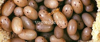
Reproduction occurs only sexually. Earthworms are hermaphrodites. Their reproductive system is located in several segments of the anterior part. The testes lie in front of the ovaries. When mating, the sperm of each of the two worms is transferred to the seminal receptacles (special cavities) of the other. Cross fertilization of worms.
During copulation (mating) and oviposition, girdle cells on the 32-37 segment secrete mucus, which serves to form an egg cocoon, and a protein liquid to nourish the developing embryo. The secretions of the girdle form a kind of mucous coupling (1).
The worm crawls out of it with its back end first, laying eggs in the mucus. The edges of the coupling stick together and a cocoon is formed, which remains in the earthen hole (2). Embryonic development of eggs occurs in a cocoon, from which young worms emerge (3).
Sense organs
The sense organs are very poorly developed. The earthworm does not have real organs of vision; their role is played by individual light-sensitive cells located in the skin. The receptors for touch, taste, and smell are also located there. Earthworms are capable of regeneration (easily restore the back part).
Text of the book “Earthworms for increasing yield”
Structure and main functions of earthworms
Body structure is the basis of knowledge about animals.
Whether we want to understand the diversity of forms of a group of animals that interests us, or to get acquainted with their way of life, their connection with their environment, or to approach the solution of certain practical issues related to these animals, the question of body structure is the main prerequisite for solving any other . In particular, as for earthworms, in order to determine the genus and species of any of their representatives, it is not enough to know its external characteristics, but it is necessary to establish a number of structural features of the internal organs by dissection. Lumbricus terrestris (side view):
1 – head blade; 2 – belt; 3 – male genital opening
In the body of an earthworm, one can distinguish the anterior (or head) end of the body, which is thicker, with stronger muscles and usually darker in color, and the posterior (or tail), thinner and paler. The back end of the worm is often flat. The mouth is located at the head end of the body, and the anus is located at the tail end. The dorsal side, which is more convex and usually darker, and the ventral side, which is lighter and flatter, also differ well from each other. In worms preserved in alcohol or formaldehyde, the ventral side may be concave in places or along the entire length.
The entire body of an earthworm is divided by transverse constrictions into separate sections called segments or segments. This ringing, or segmentation, is the leading feature of their organization: each of the segments, in principle, has the same structure and contains basically the entire complex of organs characteristic of these animals. In the anterior part of the body the segments are larger; towards the rear their size gradually decreases.
The number of segments in common species varies from 90 to 300; it is subject to significant fluctuations in different specimens of the same species, but, unlike many of their aquatic relatives, it does not change with age. Only in some tropical species the number of segments reaches 600.
Taking a close look at the surface of the body, you can see that each segment is divided into three parts by two shallow grooves. This is the so-called secondary ringing, which also reflects some features of the internal organization of each segment. The body segments are numbered, with the head segment being considered the first.
Head blade shapes:
a – epilobic; b – tanilobic
In earthworms, the head segment can be of two types: either the head lobe, protruding on the dorsal side into the region of the first segment, is separated from it by a transverse groove, or it reaches the groove between the first and second segments. In the first case, the head segment is called epilobic, in the second – tanilobic. These differences in the shape of the head lobe are important in identifying worm species. The head lobe is an organ of touch and smell; With it, the worm examines objects encountered on its way.
In the anterior part of the body in adult individuals there is a so-called girdle, X - a thickening covering from 5 to 12 segments, usually differently colored compared to the rest of the body. The skin in the girdle area contains a large number of glands that secrete nutrients for eggs when egg cocoons are laid. Therefore, during the breeding season, the girdle looks very swollen, and when there are no cocoons being laid, the region of the girdle differs from neighboring areas only in color and a different character of the body surface. The shape of the girdle can be ring-shaped, if it is developed equally strongly on all sides, or saddle-shaped, if it is poorly developed on the ventral side.
On the sides of the ventral side of the girdle there are elongated thickenings, which are called maturity ridges. In some species these ridges are replaced by several pairs of mature tubercles. The shape, length, color and location of the girdle, ridges and tubercles serve as significant species characteristics of earthworms.
The anterior section of the body of the earthworm Lumbricus terrestris from the dorsal side:
1 – dorsal pores; 2 – belt
Along the entire length of the worm's body you can see small bristles, which are clearly visible through a magnifying glass. They are found on all body segments except the first. In fauna earthworms, the setae are arranged eight on each segment, in pairs or singly. The bristles form four longitudinal rows on each side of the worm’s body, which are usually designated by the letters of the Latin alphabet - a, b, c, d. Their location is of great importance in identifying worms. The rows of setae a and b, c and d are usually close together in pairs. The degree of their convergence varies among different species. When identifying worms, the ratio of the distances between the rows of bristles must also be taken into account. These distances are denoted by the letters aa, ab, be, cd and dd (as is customary to denote line segments in geometry). The ratio of the distances between the bristles to the size of the outer contour of the cross-section through the worm is also important.
The bristles are important organs of movement: the worm can cling to soil particles with them or be repelled by them when moving in soil burrows and on the surface of the earth. You can also verify their presence by running your finger along the ventral side of the body from the tail end to the head. If a live worm is placed on a sheet of paper, a characteristic rustling sound will be clearly audible as it moves, caused by the friction of the hard bristles. On some segments, the setae are modified into special sex setae, which are important during the mating of worms.
The genital openings are located on the ventral side of the body, in front of the girdle. This includes a pair of male genital pores, usually located on elevations - the so-called glandular cushions, and a pair of female genital pores, poorly distinguishable from the outside.
Diagram of the types of arrangement of bristles in cross sections through segments:
1 – bristles not close together in pairs; 2 – setae weakly close together in pairs; 3 – bristles very close together in pairs; a – ventral setae; b – abdominal-lateral; c – dorsal-lateral; d – dorsal; aa – abdominal interval; ab – abdominal-lateral; bс – lateral; cd – dorsal-lateral; dd – dorsal
In addition, most species have 2–3 pairs of seminal receptacle pores. The meaning of all these holes will be discussed below.
On the dorsal side of preserved worms, dorsal pores are clearly visible in the intersegmental grooves, the anterior border of which is important in determining the types of worms.
The body color of earthworms depends, on the one hand, on the color of their blood, on the other, on skin pigments. It is necessary to strictly distinguish between the body color of worms, which can only be discussed in relation to living individuals and which depends on the combination of skin pigment and blood color, from skin pigmentation, which is determined only by the presence of pigments. Worms lacking pigment have a pink or red body color during life, and when preserved they become white or grayish; pigmented species can be red, brown, brown, yellow and blue.
The body length of earthworms ranges from 2 to 30 cm with a thickness of 2 to 12 mm. In tropical countries there are species that reach a length of 3 m. The bulk of worms inhabiting soils around the world are represented by species that are 5–20 cm in length.
All further presentation refers to earthworms of the Lumbricidae family.
Worms of other families (except for botanical gardens, where worms are sometimes brought along with tropical plants) can only be found in the Ussuri region, Central Asia and the southern part of the Black Sea coast of the Caucasus. Intestinal structure and digestive function
The mouth, located at the anterior end of the body, leads into a small oral cavity with folded walls, followed by a muscular pharynx. Due to the fact that the pharynx is connected by a complex interweaving of muscle fibers to the body wall, it not only makes swallowing movements and compresses ingested substances, but can also turn out through a wide open mouth. These movements allow the grasping of objects such as leaves, pebbles, etc., used for food or for other purposes.
In the thickness of the pharyngeal wall and beyond there are numerous pharyngeal glands, the ducts of which open directly into the pharynx or into a special pocket in the dorsal thickened part of its wall. The pharyngeal glands secrete a mucous fluid that envelops ingested food particles. In this respect, their function is similar to that of the salivary glands in other animals. But, in addition, the pharyngeal glands produce a substance that digests proteins; it is active in an alkaline environment and is similar in its action to the enzyme that enters the intestines from the pancreas in vertebrates. Thus, the chemical processing of proteins begins in earthworms already in the oral cavity, which is probably due to the need for the most complete extraction of protein substances from food (as a rule, extremely poor in these substances).
Diagram of the structure of the anterior end of the earthworm's body:
1 – suprapharyngeal ganglion; 2 – mouth; 3 – pharynx; 4 – abdominal nerve cord: 5 – esophagus with openings of calcareous glands in segments 9–11 and 13; 6 – goiter; 7 – muscular stomach; 8 – midgut; 9 – abdominal; 10 – dorsal vessel; 11 – skin epithelium; 12 – muscles of the body wall; 13 – places of branches of the annular vessels from the spinal vessel in segments 7–11; 14 – pharyngeal pocket (reproductive system not shown)
The pharynx passes into the esophagus. This is a rather narrow cylindrical tube, the walls of which have well-developed muscles. On the sides of the esophagus there are 1-3 pairs of lateral pockets - the so-called calcareous glands. In some species they are located deep in the wall of the esophagus and are therefore invisible from the outside. These glands are called calcareous due to the fact that crystals of lime carbonate are found in them under a microscope. The fact that these glands produce lime is proven by the fact that food masses are significantly enriched with it as they pass through the intestines (the amount of lime carbonate in the intestinal contents can increase from 0.8 to 1.3–1.8%). It was assumed that the role of these glands is to neutralize the acids contained in the ingested soil. This assumption is based on good agreement with the above-mentioned need for an alkaline environment for the activity of digestive enzymes. However, this hardly exhausts the role of calcareous glands. There are many other assumptions regarding their function, and the most varied ones; This already shows that the function of the calcareous glands must be considered unclear.
Behind the esophagus there is a voluminous expansion of the intestinal tube - the so-called goiter, occupying 2-3 segments. Ingested food accumulates in it, which from there enters in portions into the following sections of the intestine. In the absence of such a device, the body would not have time to cope with the processing of incoming material. The goiter has fairly thin elastic walls, due to which it stretches well.
Directly behind the goiter there is another extension of the intestinal tube - the muscular stomach. Inside, it is lined with epithelium with a thick cuticle, and its wall consists of annular and longitudinal layers of muscles, with the inner annular layer having a “feathery” structure, similar to the longitudinal layer of muscles of the body wall, being especially well developed. The task of the stomach is to grind food; In this process, the main role is played, just as in chickens and other granivorous birds, by the friction of mineral soil particles against each other, between which there are organic food substances. Darwin observed that grains of sand and pieces of brick that passed through the intestines of earthworms took on a rounded shape instead of an angular one. There are new observations and experiments proving the importance of mineral soil particles for grinding food in the intestines of worms; in their absence (for example, if the worms are placed in peat), they starve, despite the abundant food in the form of leaves.
The gizzard is followed by the midgut, which extends to the posterior end of the body.
Cross section through the dorsal intestine of an earthworm with typhlozol (from Stolte):
1 – chloragogenic tissue; 2 – longitudinal muscles of the intestine; 3 – ring muscles of the intestine; 4 – intestinal epithelium; 5 – blood vessel; 6 – small blood vessels; 7 – dorsal blood vessel; 8 – ring vessels
A deep dorsal fold, or typhlozol, stretches along the entire length of the midgut, thanks to which in transverse sections the contour of the intestinal cavity takes on a horseshoe-shaped outline. The physiological significance of this peculiar feature of the organization of the intestine is clear: in this way, an increase in the absorption surface of the intestine is achieved.
The intestinal wall contains a large number of glandular cells that produce mucous secretions and digestive enzymes. Among the latter, as in the pharynx, there are enzymes that digest proteins and, in addition, enzymes that convert starch into sugars (maltose and glucose); Fats are also converted into a soluble state in the intestines.
Thus, in worms, as in vertebrates, nutrients in the form of solutions are absorbed by the intestinal wall. The movement of food is accomplished by the action of the intestinal muscles, which consists of an inner circular and outer longitudinal layer of muscle (note that the arrangement of the layers here is the opposite of that in the body wall). Some species have several layers of muscle in the intestinal wall.
In the last 10–15 segments of the body, the intestine lacks a dorsal fold, and its epithelium acquires cilia. This part is called the hindgut. Absorption no longer occurs in it, but only the process of formation of lumps of feces, i.e., those coprolites that are so important for the soil structure, takes place. On the last segment of the body, the intestine opens outwards with an anal opening, which looks like a vertical slit.
An interesting debate between two famous naturalists of the last century on the issue of earthworm food is Etienne Claparède (France), an excellent expert on invertebrates (in particular, annelids), and Charles Darwin (England). Claparède found in different parts of the intestines of earthworms the remains of crushed leaves mixed with soil, and on this basis he believed that the worms swallow the soil only for the purpose of making the plant remains they swallowed better grindable. Darwin, without denying that worms feed on fallen leaves and other plant debris, at the same time argued that they also use the ingested soil for nutrition. He observed that places where they could only feed on soil rich in organic matter (for example, neatly swept yards) were also abundantly populated by worms. All further studies confirmed the correctness of Darwin's observations.
The amount of earth absorbed and processed in the intestines of earthworms is of great importance. It turned out to be huge: by weighing the coprolites, it was established that the worms inhabiting cultivated soils pass through their intestines in 24 hours an amount of soil equal to their body weight.
Mention should also be made of the characteristic tissue that covers the outside of the entire midgut and dorsal blood vessel and fills the dorsal fold of the intestine. When opening a live or just killed earthworm, the yellow color and loose velvety surface of the midgut, on which red blood vessels stand out in contrast, attract attention. This tissue is called chloragogenous, or yellow. Its connection with the intestine is purely topographical: it is a modified part of the lining of the body cavity (peritoneal epithelium) adjacent to the intestine.
Yellow tissue consists of large cells, the plasma of which is filled with droplets of substances that have a yellowish color. The origin and nature of this substance, and at the same time the function of the tissue itself, are not entirely clear. Some researchers consider this tissue to be a storage site for reserve nutritional materials, similar to the adipose tissue of vertebrates. Indeed, inclusions of yellow tissue cells contain fat, protein and a substance similar to glycogen (animal starch). At the same time, it is known that this tissue contains large amounts of uric acid, that foreign substances introduced in the form of solutions into the body cavity (paint) accumulate in the cells of chloragogenous tissue, and that the final nitrogenous metabolic products to be excreted from the body are usually have a yellow or brown color. All this makes us think about the excretory function of this tissue.
It is very likely that, along with the accumulation of reserve nutrients, the cells of the yellow tissue have the ability to extract waste products formed during the metabolic process from the blood circulating in it and the fluid filling the body cavity.
Once inside the cells of the yellow tissue, these substances are switched off from the blood flow and become harmless. Gradually accumulating in the cells of this tissue, they can remain there for a long time. But they can also be excreted from the body, since the cells of the yellow tissue often break off and enter the body cavity, and from there they are brought out along with the splashing of the cavity fluid through the dorsal pores. Excretory organs
The excretory function is performed in earthworms (as in all ringworms) by tubular organs located in pairs in each segment, except the anterior ones. These organs are called nephridia, which means “kidney-like organ” in Greek. Nephridia are located in the body cavity on the sides of the intestines. Each of them is a convoluted tube starting inside the body with an opening into the body cavity located on the capitate extension, the cells of which are equipped with cilia. This expansion is called, by analogy with similar formations in more primitive rings, a funnel. Almost immediately behind the funnel, the nephridium pierces the intersegmental septum and penetrates into the next segment of the body.
There it first forms a highly convoluted thin tube, which passes into a wider middle part of the nephridium, equipped with cilia. Then the nephridium, making several loops, passes into the excretory part, which ends on the ventral side of the body with an external opening, or nephridial pore. It is very difficult to find it from the outside, since its edges are always tightly closed. Not far from the nephridial pore there is an extension of the nephridial tube, which is something like a bladder. Nephridia are equipped with a very rich network of blood vessels. The blood leaving the nephridium enters the transverse vessel, and from it into the dorsal vessel.
The structure of the nephridium of an earthworm:
1 – funnel; 2 – dissemination; 3 – convoluted part of the nephridium tube; 4 – ciliated part of the nephridium tube; 5 – “bladder”; 6 – time of nephridium; 7 – outer loop
It should be noted that in one of the earthworms (Allolobophora autipae), the nephridial tubes do not open with pores independent from each other, and their outer parts flow into longitudinal excretory canals, which run to the right and left along the entire body and at its posterior end flow into the intestine not far away from the anus. Thus, here the connection of the excretory apparatus into one anatomical whole is outlined and a connection is established with the hind intestine.
The cells of the thin part of the nephridial tube capture nitrogen metabolism products from the blood circulating outside the network of nephridial capillaries to be excreted from the body.
These substances enter the cavity of the nephridial tube and here they mix with the cavity fluid entering through the funnel at the inner end of the nephridium. The cavity fluid also contains excretory products, dead cells, worn-out bristles, etc. The fluid of the nephridial tube is driven by the beating of cilia towards the excretory end, from where it is periodically released through the external pore by contraction of the muscles of the body wall (Roots, 1955).
There is evidence that the terminal nephridium vesicle is emptied once every three days. Other observations indicate that a worm weighing 1–1.8 g secretes 0.82 cm of excrement per day. Such quantities must be excreted from the body several times a day.
The excreta contains, in general, the same substances as in mammals, namely: urea, ammonia, creatinine, salts, etc., but in much lower concentrations. However, normal excrement of worms contains 0.3% protein, while in higher animals there is no protein in the excretory products.
The cells of the middle part of the nephridial tube have the ability to phagocytose, i.e., to actively ingest water-insoluble substances from the body cavity (dead cells, coagulated protein, bacteria, etc.). These substances accumulate there for an indefinite period of time. Sanitary services of this kind are also performed by other cells within the body: amoeboid blood cells, cells of the body cavity and the aforementioned cells of chloragogenous, or yellow, tissue.
Especially many amoeboid cells, swallowed foreign bodies, are found in the body cavity. They get here by actively crawling out of the vessels, squeezing between the cells of the vascular wall. These cells are removed from the body cavity in different ways. Firstly, they crawl through the intestinal wall and, entering its cavity, are excreted along with feces (this has been observed many times). Secondly, as already mentioned, they can exit with the cavity fluid through the nephridia. And, thirdly, they can come out along with the cavity fluid sprayed through the dorsal pores.
Blood vessels of nephridium earthworm:
1 – funnel; 2 – abdominal blood vessel; 3 – dissemination; 4 – abdominal nerve cord; 5 – subnervous vessel; 6 – nephridium tube with blood vessels
In general, it can be assumed that the cavity fluid is replaced quite quickly. This is why it becomes so important in worms during the process of excretion.
Water continuously enters the body of the worms and is released back into the external environment in the ways indicated above. Thus, the body of the worm and, in particular, the body cavity is constantly rinsed with water. Therefore, for the normal functioning of these physiological functions, worms need environmental conditions that would ensure that water enters their body in much greater quantities than most terrestrial animals.
How does water enter the body of worms?
It should be noted that worms never drink. They absorb water over the entire surface of their body; water passes through the integument and muscles, accumulating in the body cavity. In this case, worms can only use water in a liquid state. A worm in an environment containing water vapor may die from drying out if it has no other source of moisture.
Under normal conditions, the body of worms contains about 84% water. Despite such a significant supply of water, this is far from the limit. If the worm is given the opportunity to further increase the supply of water in its body, it will immediately do so. This is easy to verify if you put earthworms in water. After a few hours, their weight will increase significantly due to the water absorbed by the surface of the body. After being removed from the water, the worm returns to its original weight, and this happens in a very short time (1–2 hours). Removal of excess water from the body occurs in a very unique way: it is absorbed by the intestinal cells, from them enters the cavity of the latter and is removed mainly through the anus, and partly through the mouth.
Under normal living conditions in the soil, the function of removing excess water is performed by nephridia. The presence of a constant flow of water through the body by absorption by the surface of the body and excretion of excess by the kidneys is a very common phenomenon among aquatic animals. It is undoubtedly inherited by earthworms from their aquatic ancestors.
The metabolism of aquatic animals occurs with increased circulation of water through their body. They cannot be threatened by a lack of water, whereas in soil conditions with this type of water exchange, a sufficient amount of moisture becomes the main factor ensuring the possibility of existence. Therefore, soil moisture is important when colonizing them with certain types of earthworms.
It is also known about the ability of earthworms to lose water during periods of drought and wintering, which is associated with their transition to a state of hidden life.
Germ layers
The germ layers are the basis of all organs. In annelids, the ectoderm (outer layer of cells), endoderm (inner layer of cells) and mesoderm (intermediate layer of cells) appear early in development as three germ layers. They give rise to all major organ systems, including the secondary cavity and the circulatory system.
These same organ systems are subsequently preserved in all higher animals, and they are formed from the same three germ layers. Thus, higher animals in their development repeat the evolutionary development of their ancestors.
Earthworm
belongs to the group of annelids. It does not have any special organs specifically designed for gas exchange, and gas exchange occurs by diffusion across the entire surface of the body. In essence, they do not need specialized organs, since, due to the cylindrical shape of the body, their surface area to volume ratio is large, and with their relatively low activity, they do not consume much oxygen.
However, in annelids
there is a circulatory system (unlike some simpler animals and single-celled organisms), and the respiratory pigment hemoglobin is dissolved in their blood. Contractions of large blood vessels drive blood along with gases dissolved in it throughout the body; this also contributes to the maintenance of steep diffusion gradients.
Thin skin of an earthworm
(cuticle) is constantly moistened by the secretion of the glands located in the epithelium.
Capillaries are located in the epithelium directly under the cuticle. The distance between the blood vessels and the surface of the body is small and this ensures rapid diffusion of oxygen into the blood. Earthworms are practically not protected from drying out and therefore try to stay only in a humid environment. A. Tracheal system in locusts. B. The structure of the insect trachea.
Respiratory system of insects - locusts.
Gas exchange in insects
carried out through a system of tubes, the so-called trachea. This system allows oxygen to flow from the air directly to the tissues and there is no need for its transportation by blood. This is a much faster method than diffusion of dissolved oxygen through tissue; Such gas exchange creates conditions for high metabolic rate.
Spiracles
— paired openings located on the second and third thoracic and on the first eight abdominal segments of the insect’s body lead to air cavities. Branched tubes - tracheas - extend from these cavities. Each trachea is lined by an epithelium that secretes a thin layer of chitinous material. Typically, this rigid layer is further reinforced by spiral and annular thickenings, due to which the airways remain open even if the pressure in the lumen of the trachea is negative (compare with the cartilaginous rings in the human trachea and bronchi). At each body segment, the trachea branches into numerous smaller tubes called tracheoles; tracheoles also branch, penetrating the tissues of the insect, and in the most active tissues, for example in the flight muscles, they end blindly inside individual cells. The degree of branching of tracheoles can vary depending on the metabolic needs of the tissues.
Tracheoles have chitinous lining
absent. At rest they are filled with watery fluid; at this time, oxygen diffuses through them to the tissues (and CO 2 in the opposite direction) at a speed quite sufficient to satisfy the needs of the insect. In the active state, increased metabolic activity of the muscles leads to the accumulation of certain metabolites, in particular lactic acid, and the osmotic pressure in the tissues increases accordingly. When this happens, the fluid from the tracheoles, under the influence of osmotic forces, is partially absorbed into the tissues, and more air enters the tracheoles, and therefore more oxygen, and this oxygen is supplied directly to the tissues just when they need it.
Conditions created in insect tissues at rest and in an active state (work of tracheoles).
The overall flow of air passing through the insect's body is regulated by a mechanism that closes the spiracles
. The opening of each spiracle is equipped with a system of valves controlled by very small muscles. The edges of this opening are covered with hairs, which prevent foreign particles from entering the spiracles and prevent excessive loss of moisture. The size of the hole is adjusted depending on the amount of CO 2 in the insect’s body.
Increased activity leads to increased formation of CO 2. Chemoreceptors
they catch this and the spiracles open. The same stimulus can also cause ventilatory movements of the body, especially in large insects such as locusts. The dorsoventral muscles, contracting, make the insect's body flatter, as a result of which the volume of the tracheal system decreases and air is pushed out of it (“exhalation”). Air absorption (“inhalation”) occurs passively when the body segments, due to their elasticity, return to their original shape.
Judging by some data, the thoracic and abdominal spiracles
open and close alternately, and this, combined with the ventilation movements of the body, creates a unidirectional flow of air that enters the insect's body through the thoracic region and exits through the abdominal region.
Tracheal system
, of course, is very effective in terms of gas exchange, however, it should be borne in mind that gas exchange in most insects is determined exclusively by the diffusion of oxygen through the tissues of the insect. Diffusion, as is known, is effective only over short distances, and this imposes severe restrictions on the size that insects can reach. These small distances at which diffusion is quite effective do not exceed 1 cm; therefore, although there are insects up to 30 cm long, their body should not be more than 2 cm thick.